


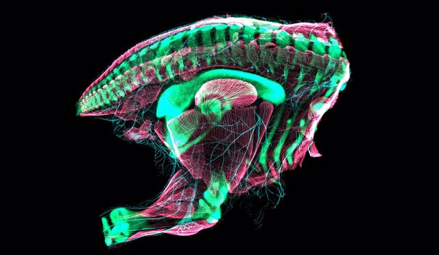
Embryonic quail hindquarters imaged by confocal laser scanning microscopy. The pelvis of this quail embryo has just transformed into a relatively “modern” bird configuration. – CHRISTOPHER T. GRIFFIN AND BHART-ANJAN S. BHULLAR
Aug. 5 () –
All birds in gestation have a moment before breaking the egg shell in which the hip bone is a small replica of a dinosaur’s pelvis.
That’s one of the findings of a new Yale-led study in the journal Nature exploring the evolutionary underpinnings of the avian hip bone. It’s also a nod to the dramatic transformation it took from dinosaurs to birds over tens of millions of years.
“Every bird, in its early life, has this dinosaur form,” he said. it’s a statement Bhart-Anjan S. Bhullar, assistant professor of Earth and planetary sciences at Yale and lead and corresponding author of the new study. “Then at the last minute, it’s like he remembers he’s a bird and he needs a bird’s pelvis.”
Over the past decade, Bhullar and his collaborators have conducted groundbreaking research on key evolutionary transitions between dinosaur, reptile, and bird species, including the development of the inner ear of dinosaurs, the beak of birds, the movable jaw of mammals and sight in vertebrates.
Bhullar’s lab is particularly known for its innovative use of computed tomography (CT) and microscopy to create 3D images of animal embryos.
Christopher Griffin, a postdoctoral associate in Bhullar’s lab, is the lead author of the study. He and Bhullar, with their colleagues, observed pelvic development in alligators, domestic chickens, Japanese quail, Chilean tinamou, and parakeets, and compared their developmental stages to those of dinosaurs, including the feathered species Archeopteryx.
For the study, the team tagged embryonic hip bones with antibodies to look for proteins that are expressed in developing cartilage, connective tissue, skeletal muscles and nerves. The researchers created 3D images of the hip bones, muscles and nerves using confocal microscopes and CT scans.

They found that the avian pelvis is an example of “terminal addition,” a biological mechanism in which ancestral features appear in an animal until late in its development. This was a surprise, Griffin noted, because many important features in the transition from dinosaur to bird, such as the bird’s beak, they are seen early in a bird’s embryonic development.
“It was unexpected to find that these early stages of bird development looked so much like the hips of an early dinosaur,” Griffin said. “Over just two days, the developing embryo changes in a way that mirrors how it changed in evolution, going from resembling a primitive dinosaur to resembling a modern bird”.
The hip bone is the core of a bird’s body. It runs the length of the avian frame, wrapping around the torso, while also allowing a bird to stand, move and carry its full body weight.
“The bird’s body is incredibly modified in virtually every way to create an optimized flying machine,” Bhullar explained. “Their body structures are heavily constrained by the needs of aircraft design.”
The new study also looked at the birds’ muscles and nerves related to hip development. The researchers said that the development of those systems was out of sync with bone development, implying that each system was somewhat “decoupled” from the others.


Add Comment